 |
|
|
|
|
 Back to 1997 1st Quarter Table of Contents
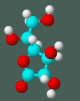
When there is concern about a patient’s fat intake or blood lipid levels, tests ordered are usually restricted to cholesterol, triglyceride, HDL, and LDL. Occasionally, a lipoprotein electrophoresis or lipoprotein-a (Lp-a) are ordered. In addition to those tests listed above, at The Center we routinely measure red blood cell membrane (RBC) fatty acids using modifications of existing procedures.1-3 RBC membrane fatty acid analysis is a very difficult procedure to perform and The Center is one of the few clinical institutions in the U.S.A. performing this test. The test is very complex and involves lipid extraction, methylation, separation steps and final analysis by gas chromatography. Plasma fatty acids can also be measured. However, the RBC profile is preferred because RBC fatty acids reveal long-term fatty acid balance in the tissues and is not influenced by recent dietary fat intake. Fatty acids are the most simple molecular form of dietary fats and may be saturated (no double bonds, or unsaturated (one or more double bonds). Three of the fatty acids cannot be made by the body and must be obtained through the diet. These essential fatty acids are linoleic (18 carbons with two unsaturated bonds), alpha linolenic (18 carbons and three unsaturated bonds), and arachidonic (20 carbons and four unsaturated bonds). All the essential fatty acids are, by definition, polyunsaturated. Fatty acids of greatest importance to human nutrition are long chains of 12 to 20 even numbered carbons. In the body, fatty acids serve as energy sources, precursors of prostaglandins, components of cell membranes and myelinization of the CNS. Fatty acids are identified by common names (linoleic, arachidonic); by chemical structure (C18:2n-6, C20:4n-6); or by the Greek terminology, omega, referring to the location of the first double bond from the end carbon of the fatty acid chain. Omega is the last letter in the Greek alphabet and designates the last carbon in the chain, therefore, fatty acids may be classified as either omega-3 (third carbon), omega-6 (sixth carbon) or omega-9 (Table 1, Page 21) A stearic/oleic ratio (SA/OA) is included in the RBC fatty acid profile. Apostolov, et al, reported the SA/OA ratio was lower in RBC membranes from cancer patients than RBCs from patients without cancer.4-6 Other investigators have suggested that the SA/OA ratio can be used as a tumor marker.7-10 A ratio of less than 1 may be suggestive of cancer, while a ratio of less than 0.7 is said to be a marker for cancer. Unpublished work from The Center’s RECNAC cancer research unit (RECNAC is cancer spelled backwards), confirms this finding in cell culture. When the SA/OA ratio was measured in the membranes of four normal cell lines, the ratio was always greater than 0.7. When the SA/OA ratio was measured in seven cancer cell lines, the ratio in all cancer cells was less than 0.7 (p<0.05 for every pair of normal and cancer cell lines). There are various theories why the ratio change may indicate the presence of cancer. These range from changing the fluidity of the membrane (saturated fatty acid, stearic, to unsaturated fatty acid, oleic), or by causing differences in material exchange through the membrane.11 Red Blood Cell Membrane Fatty Acids as a Diagnostic Test Table 1. Commonly Occurring Fatty Acids* Number of Number of Omega Name Carbons Double Bonds Classification SaturatedLauric 12 0 None Myristic 14 0 None Palmitic 16 0 None Stearic 18 0 None Arachidic 20 0 None MonounsaturatedPalmitoleic 16 1 Omega-9Oleic 18 1 Omega-9 PolyunsaturatedLinoleic* 18 2 Omega-6 Linolenic* 18 3 Omega-3 (alpha)+ Arachidonic* 20 4 Omega-6 * Partial list - indicates essential fatty acids + Gamma linolenic (GLA) is an Omega-6 fatty acid Although our research data with cell culture lines confirmed the low SA/OA ratio in cancer cells, in patients with cancer followed at The Center, the patient’s SA/OA ratio rarely is less than 0.7 unless the cancer is in an advanced state. In ten patients with cancer randomly selected from our files (ages14 to 66), the mean SA/OA ratio was 1.12. What clinical information can be obtained from measuring RBC membrane fatty acids? The analysis, including ratios and various fatty acid metabolites, can be helpful in prevention and treatment of heart disease, skin disorders, arthritis, hypertension, reproductive disorders, cancer, and imbalances that relate to degenerative diseases, depression of the immune system, and production of inflammatory prostaglandins. Since fatty acids function relative to each other, a correct balance of essential and nonessential fatty acids is necessary for good health. The RBC membrane fatty acid profile performed at The Center is shown in Table 2 (page 22). An example of the clinical use of the RBC membrane fatty acid profile is shown in the following patient, a 59 year old female. When first seen at The Center, she had a history of diabetes with retinopathy, allergies, carpal tunnel syndrome and yeast infection. Abnormal RBC membrane fatty acid profiles were correlated with these pathologies. She was low in three out of four omega-6 fatty acids; normal in three omega 3 fatty acids; low in oleic and high in nervonic fatty acids; high in palmitic and low in stearic fatty acids; and had a SA/OA ratio of 1.2. Since fatty acid imbalances may lead to (or exacerbate) degenerative diseases, it is important to detect and correct these imbalances as well as to treat the usual disease symptoms. Table 2 The Center’s Red Blood Cell Membrane Profile Expected Omega-6 Fatty Acids Values Linoleic (LA) 8.8-14.0 Gamma Linolenic (GLA) 0.04-0.08 Dihomogammalinolenic (DGLA) 1.03-2.23 Arachidonic (AA) 12.18-17.09 Omega-3 Fatty Acids
Stearic/Oleic Ratio 1.0 or >
By offering this test to our patients, we are able to help them early on their road to recovery and to improve their health. References
|

This website is managed by Riordan Clinic
A Non-profit 501(c)(3) Medical, Research and Educational Organization
3100 North Hillside Avenue, Wichita, KS 67219 USA
Phone: 316-682-3100; Fax: 316-682-5054
© (Riordan Clinic) 2004 - 2024c
Information on Orthomolecular.org is provided for educational purposes only. It is not intended as medical advice.
Consult your orthomolecular health care professional for individual guidance on specific health problems.
 Download The Full Text Article in (PDF)